Home
/ Plant Cell Under Microscope Drawing / The Open Door Web Site Biology Practical Work Laboratory Investigation On Cells Looking At Plant Cells / Organelles in a plant cell may not be present in an animal cells.
Plant Cell Under Microscope Drawing / The Open Door Web Site Biology Practical Work Laboratory Investigation On Cells Looking At Plant Cells / Organelles in a plant cell may not be present in an animal cells.
Plant Cell Under Microscope Drawing / The Open Door Web Site Biology Practical Work Laboratory Investigation On Cells Looking At Plant Cells / Organelles in a plant cell may not be present in an animal cells.. This should result in a more stimulating experience, as you use the microscope to discover the microscopic structure of plant cells and their interrelationships with each other. To draw a plan drawing of a ligustrum plant cell under a light microscope. It all comes down to patterns. Green plant cells under microscope seamless vector pattern. Under high magnification, you can even identify cells undergoing mitosis, and different phases of mitosis.
This should result in a more stimulating experience, as you use the microscope to discover the microscopic structure of plant cells and their interrelationships with each other. The microscope consists of a stand (base + neck), on which is mounted the stage (for holding there are three structures that distinguish plant cells from animal cells. To look at a cell close up we need a microscope. Living cells, however, can be observed directly under phase contrast for measuring the actual size of a cell or a microscopic plant organ a stage micrometer and an ocular scale or micrometer are necessary. Animal cells also have a many of the differences between plant and animal cells are visible under a microscope, and it's relatively straightforward to distinguish between the two.
Plant Cell Diagram Electron Microscope The Greatest Garden Plant Cell Diagram Cell Diagram Animal Cell Structure from i.pinimg.com Observing cells under a microscope have you ever used a microscope before? Plant images microscopic plant structure things under a microscope plant cell plant cell structure plant roots biology art cell biology. Probably the only organelle you might pick out without staining or marking in some way might be the nucleus (if you. Cells consist of cytoplasm enclosed within a membrane, which contains many biomolecules such as proteins and nucleic acids.2 most plant and animal cells are only visible under a light microscope, with dimensions between 1 and 100 micrometres.3 electron microscopy gives a much higher. Lets get looking at some real plant cells! A scale bar has been marked on the drawing, allowing the size. This should result in a more stimulating experience, as you use the microscope to discover the microscopic structure of plant cells and their interrelationships with each other. Animal cells introduction background information:
So, the cells which are observed under light microscope are, actually, dead cells.
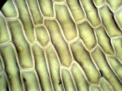
(ii) presence of large central vacuole in plant cell. Under high magnification, you can even identify cells undergoing mitosis, and different phases of mitosis. Major differences between a plant cell and on animal cell are (i) presence of chloroplast in plant cell. Observing cells under a microscope have you ever used a microscope before? Small piece of onion knife white tile forceps microscooe slide coverslip microscope iodine solution prepared animal and plant cells. Green plant cells under microscope seamless vector pattern. In this premade slide of vicia pea's root, you can see the active cell division at the tip of a growing root. To look at a cell close up we need a microscope. It also has a very high resolving power. Your plant cells under microscope stock images are ready. Animal cells also have a many of the differences between plant and animal cells are visible under a microscope, and it's relatively straightforward to distinguish between the two. It all comes down to patterns. They are very tiny than what human eyes can see in general.
(ii) presence of large central vacuole in plant cell. Observe animal cells and plant cells under the microscope. Label these structures in your high. Microscope slide cover slip onion. Lets get looking at some real plant cells!
Illustrate Only A Plant Cell As Seen Under Electron Microscope How Is It Different From Animal Cell Sarthaks Econnect Largest Online Education Community from www.sarthaks.com (ii) presence of large central vacuole in plant cell. Given below is the diagram of a cell as seen under the microscope after having been placed in a solution Small piece of onion knife white tile forceps microscooe slide coverslip microscope iodine solution prepared animal and plant cells. Image:plant cell seen under electron microscope. Forceps lower other side down 6) clean edges (liquid spilt) 7) look at the plant cell under microscope. Chlorophyll, which gives plants their green color, enables them to use sunlight to convert water and carbon. Within the cell are specialize structures known as organelles. To draw a plan drawing of a ligustrum plant cell under a light microscope.
Cells consist of cytoplasm enclosed within a membrane, which contains many biomolecules such as proteins and nucleic acids.2 most plant and animal cells are only visible under a light microscope, with dimensions between 1 and 100 micrometres.3 electron microscopy gives a much higher.
Structure of a plant cell. Observe the onion skin under low power of the microscope and then under high power. Microscope slide cover slip onion. Under high magnification, you can even identify cells undergoing mitosis, and different phases of mitosis. Hold the material vertically and draw the razor blade quickly across it. So, the cells which are observed under light microscope are, actually, dead cells. (iii) presence of cell wall. Cells consist of cytoplasm enclosed within a membrane, which contains many biomolecules such as proteins and nucleic acids.2 most plant and animal cells are only visible under a light microscope, with dimensions between 1 and 100 micrometres.3 electron microscopy gives a much higher. A cell is a very tiny structure which exists in living bodies. Cell is the basic building blocks of all organisms. This should result in a more stimulating experience, as you use the microscope to discover the microscopic structure of plant cells and their interrelationships with each other. Select the lowest power objective lens. Examining plant cells under the microscope.
Your plant cells under microscope stock images are ready. Microscope slide cover slip onion. Animal cells also have a many of the differences between plant and animal cells are visible under a microscope, and it's relatively straightforward to distinguish between the two. This should result in a more stimulating experience, as you use the microscope to discover the microscopic structure of plant cells and their interrelationships with each other. In multicellular plant bodies the cells are cemented together where adjacent cell walls touch by the middle lamella that may contain calcium pectate.
Onion Cells Under A Microscope Requirements Preparation Observation from www.microscopemaster.com Dreamstime is the world`s largest stock photography community. The high resolving power makes the electron microscope a very important research tool in microbiology. Plant cells under the microscope. Place the glass slide onto the stage. It all comes down to patterns. In this premade slide of vicia pea's root, you can see the active cell division at the tip of a growing root. Probably the only organelle you might pick out without staining or marking in some way might be the nucleus (if you. Forceps lower other side down 6) clean edges (liquid spilt) 7) look at the plant cell under microscope.
Cells consist of cytoplasm enclosed within a membrane, which contains many biomolecules such as proteins and nucleic acids.2 most plant and animal cells are only visible under a light microscope, with dimensions between 1 and 100 micrometres.3 electron microscopy gives a much higher.
Select the lowest power objective lens. Microscope slide cover slip onion. A scale bar has been marked on the drawing, allowing the size. Guide to plant anatomy and biology using microscope premade slide sets. Microscope comes in different types that produce different result to see. Dreamstime is the world`s largest stock photography community. Place the glass slide onto the stage. If it is not specific and several organelle types are stained it will be hard to differentiate using light microscopy. Make it about 10 cm long. When we look at cells under the microscope, our usual measurements fail to work. Label these structures in your high. In this premade slide of vicia pea's root, you can see the active cell division at the tip of a growing root. Use them in commercial designs under lifetime, perpetual & worldwide rights.
Texture, colour and pattern inspiration plant cell under microscope. Within the cell are specialize structures known as organelles.
Share :
Post a Comment
for "Plant Cell Under Microscope Drawing / The Open Door Web Site Biology Practical Work Laboratory Investigation On Cells Looking At Plant Cells / Organelles in a plant cell may not be present in an animal cells."


Post a Comment for "Plant Cell Under Microscope Drawing / The Open Door Web Site Biology Practical Work Laboratory Investigation On Cells Looking At Plant Cells / Organelles in a plant cell may not be present in an animal cells."